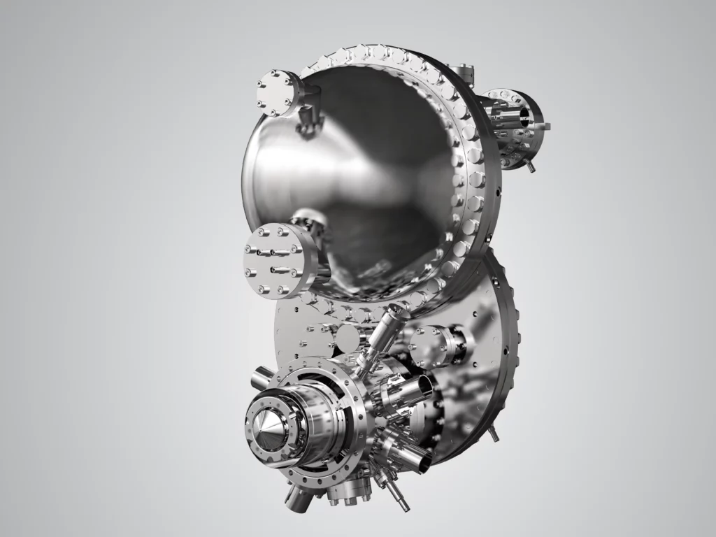
The NanoESCA is a sophisticated analytical instrument designed for imaging and small spot photoemission spectroscopy as well as µ-ARUPS.
The NanoESCA has been developed within a tight collaboration of FOCUS GmbH, Huenstetten and Scienta Omicron GmbH, Taunusstein. Its basic concept, the energy filtering by means of an Imaging Double Energy Analyser (IDEA) has been patented.
Together with its broad variety of laboratory excitation sources ranging from a high-power monochromatized Al Kα x-ray source to UV sources such as He-, Mercury arc- and Deuterium lamps or UV lasers, it is optimized for non-destructive chemical analysis of sample surfaces (Imaging ESCA / XPS), for imaging UPS and µ-ARUPS.

Next Generation Photoemission Tool for Real- and Momentum Microscopy. The core element consists of the patented IDEA (Imaging Dispersive Energy Analyser). Its double hemisphere design exhibits a real energy dispersive approach together with an unrevealed real and momentum space imaging quality
(D. Funnemann, M. Escher, European Patent EP 1 559 126 B1, US patent US 7 250 599 B2).
The aberration compensated band pass filter IDEA does not only eliminate the analyser’s spherical aberration but the tandem arrangement also largely retains the time structure of the electron signal, unlike a single hemispherical analyser which can be helpful with time resolving experiments.

Hard x-ray photoelectron spectroscopy (HAXPES) has now matured into a well-established technique as a bulk sensitive probe of the electronic structure due to the larger escape depth of the highly energetic electrons. In order to enable HAXPES studies with high lateral resolution, we have set up a dedicated energy-filtered hard x-ray photoemission electron microscope (HAXPEEM) working with electron kinetic energies up to 10 keV. It is based on the NanoESCA design and also preserves the performance of the instrument in the low and medium energy range. In this way, spectromicroscopy can be performed from threshold to hard x-ray photoemission.
Please accept YouTube cookies to play this video. By accepting you will be accessing content from YouTube, a service provided by an external third party.
If you accept this notice, your choice will be saved and the page will refresh.
The NanoESCA placed on a synchrotron beam line opens up even a few more potentials. Such sources do not only deliver just more photons. Additionally to the lab use as above the tunable wave lengths opens up the possibility to take total yield images and/or spectra for imaging resonant photo emission investigations. With the polarized synchrotron light and its pulsed time structure the NanoESCA is a beautiful tool to have a microscopic look at the magnetic dichroism of photoemission in terms of the real time behavior of microscopic magnetic domains and its spin states.
A further synchrotron related usage of the NanoESCA is the recently developed HAXPEEM. At high brilliant x-ray beam lines of synchrotrons this seems to become a highly promising approach to investigate not only surfaces with the PEEM but bulk properties of samples also.
The NanoESCA can be equipped optionally with additional electron optical elements to accomplish not only the lateral imaging but also the imaging of the angular distribution of the emitted electrons (µ-ARUPS, µ-ARPES). This concept has been called Momentum Microscope.

The Trondheim NanoESCA System
Group of Justin Wells, “quantum.wells“
Photo courtesy of Scienta Omicron
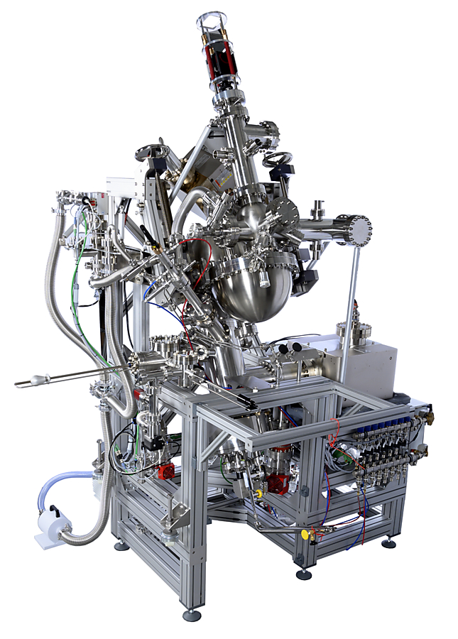
A complete NanoESCA UHV system as delivered by Scienta Omicron.
NanoESCA with 2D Imaging Spinfilter
The NanoESCA can easily be equipped with the 2D Imaging Spinfilter. The NanoESCA directly provides the real – and momentum space image at the required scattering energy of the 2D Imaging Spinfilter. This is a pre-requisite for un-compromised parallel acquisition of high resolution spin filtered images reducing measurement time by a factor of up to 10.000 compared to classical single channel detection.
The NanoESCA is distributed exclusively by our sales partner Scienta Omicron. Please contact them for more informations.
Download the Scienta Omicron Brochures here:
NanoESCA
NanoESCA II

